Orthopaedics
Cruciate Ligament Rupture: Surgery, Recovery and Realistic Timelines
Cranial cruciate ligament rupture is the most common cause of hind limb lameness in dogs, mirroring the ACL injuries seen in human athletes but usually resulting from gradual ligament degeneration rather than a single traumatic tear.
By Dr Mariana Oliveira, DVM, DACVN
4 August 2026
4 min read
Medically reviewed
Reviewed by Dr Noor Haddad, DVM, DACVD of the Dr Murphies Veterinary Review Board. Reviewed content is checked against current consensus guidance before publication.
Cranial cruciate ligament rupture is the most common cause of hind limb lameness in dogs, mirroring the ACL injuries seen in human athletes but usually resulting from gradual ligament degeneration rather than a single traumatic tear. Large-breed dogs, particularly Labradors, Rottweilers and Newfoundlands, are predisposed due to their steep tibial plateau angle, which places chronic shear stress on the ligament. Once ruptured, the tibia can shift forward relative to the femur with every step, accelerating joint damage.
Degeneration Versus a Single Traumatic Tear
The cranial cruciate ligament normally prevents the tibia from sliding forward under the femur and limits internal rotation of the joint during weight-bearing. In many dogs, chronic low-grade inflammation within the joint weakens the ligament's collagen fibres over months or years, so what looks like a sudden injury during a run is often the final failure of an already compromised structure.
Once the ligament ruptures, instability allows abnormal shearing forces across the meniscus and joint cartilage, which is why roughly half of affected dogs also suffer meniscal tears, and why the contralateral knee ruptures within one to two years in a substantial proportion of cases.
Recognising Partial and Complete Ruptures
- Sudden non-weight-bearing lameness after activity
- Sitting with the affected leg held out to the side
- Swelling on the inside of the knee joint
- Intermittent weight-bearing lameness that fluctuates over weeks
- Audible clicking suggesting an accompanying meniscal tear
Partial tears often produce a milder, intermittent lameness that owners mistake for a passing sprain, but the joint instability continues to worsen mechanically even when the dog appears to be walking reasonably well between flare-ups.
The Drawer Test and Imaging
The cranial drawer test and tibial compression test, performed with the dog relaxed or sedated, detect abnormal forward movement of the tibia relative to the femur and are the primary means of diagnosing rupture on physical examination. Radiographs help assess the degree of existing osteoarthritis and measure the tibial plateau angle, a key figure surgeons use to select the most appropriate surgical technique.
Advanced imaging such as MRI or arthroscopy is occasionally used to confirm meniscal damage before or during surgery, particularly when clinical signs suggest a torn meniscus is complicating a partial ligament tear.
Comparing Surgical Techniques
Surgical stabilisation is recommended for most dogs over 15 kilograms, since conservative management rarely restores normal function in larger, more active breeds.
- Tibial plateau levelling osteotomy (TPLO) to neutralise shear forces
- Tibial tuberosity advancement (TTA) as an alternative geometry-correcting procedure
- Lateral suture stabilisation for smaller dogs or lower-demand lifestyles
- Structured physiotherapy programme beginning within days of surgery
What Recovery Actually Looks Like Week by Week
Recovery follows a fairly predictable arc: gentle weight-bearing typically begins within a week, most dogs are walking comfortably on lead by four to six weeks, and full return to running or off-lead activity is generally not permitted until twelve to sixteen weeks post-surgery once radiographs confirm bone healing around the osteotomy. Strict activity restriction during the early weeks is critical, since premature loading can loosen implants or fracture the tibial plateau before it has fully healed.
Dogs managed with TPLO or TTA generally return to a higher level of function than those managed with lateral suture techniques, particularly in dogs over 20 kilograms, though all techniques require several months of committed rehabilitation to reach their best outcome.
When to seek help urgently
- Sudden inability to bear any weight on either hind leg
- Marked swelling or heat around a recently operated knee
- Discharge or opening of the surgical incision
- Fever or lethargy in the days following surgery
Cruciate rupture responds well to timely surgical correction, but the right technique and rehabilitation plan depend on your dog's size, activity level and joint conformation, which your vet or a referral surgeon can assess directly.
Vet-recommended
Products our review board rates for this
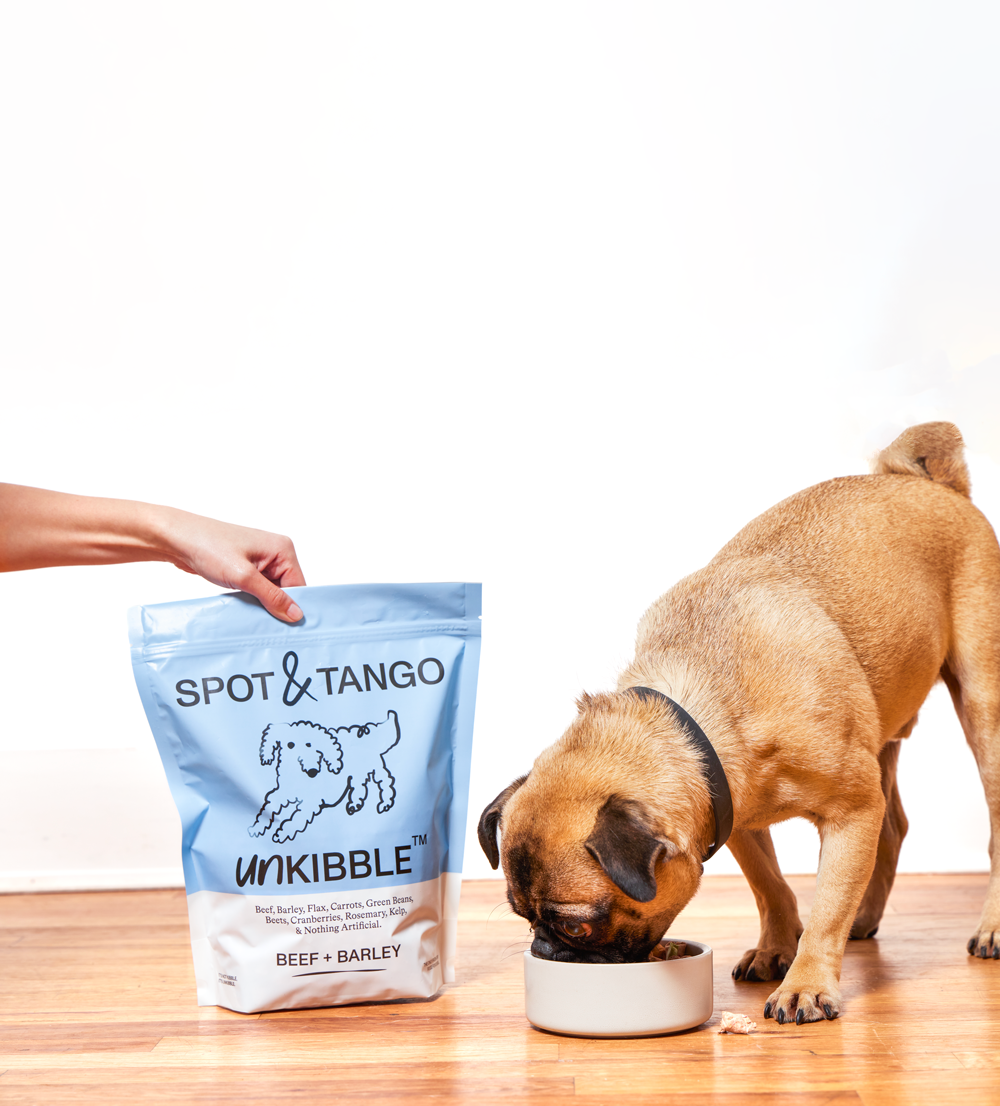
Spot & Tango
9.1Vet-formulated fresh trays and dehydrated UnKibble, portioned to the individual dog.

Lemonade Pet Insurance
9.4The lowest premiums in the category, with claims paid in minutes rather than weeks.
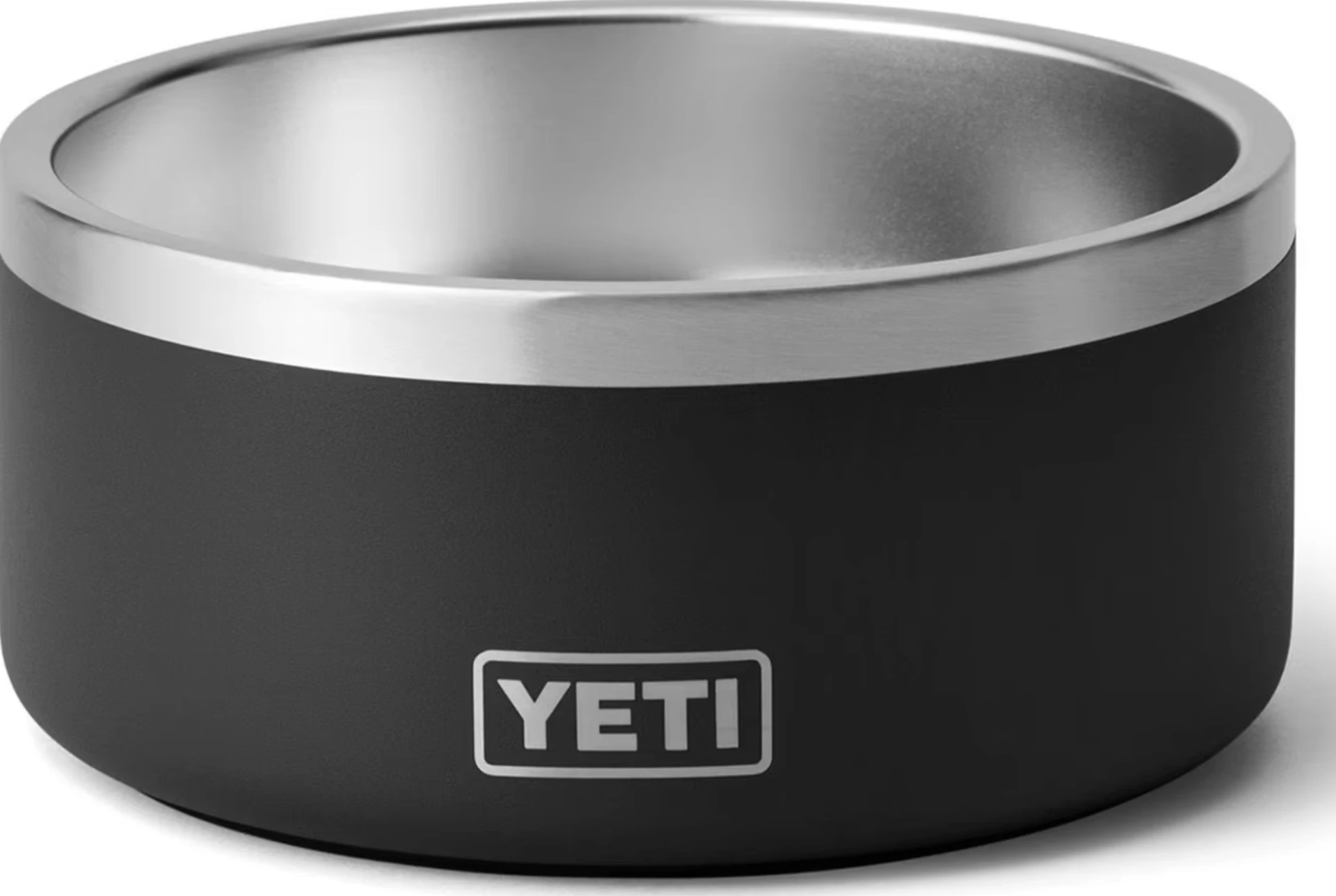
YETI Boomer 4 Dog Bowl
8.518/8 stainless, non-slip ring, dishwasher-safe — the last bowl you buy.
Recommendations are chosen by the Dr Murphies veterinary review board. Some links are affiliate links and we may earn a commission; this never affects our scoring, and brands do not see our copy before publication.
Dr Mariana Oliveira, DVM, DACVN
Veterinary nutritionist