Oncology
Mast Cell Tumours: The Great Imitator of Canine Lumps
Mast cell tumours are among the most common skin tumours in dogs and have earned a reputation as the great imitator because they can look like almost anything, from a soft, harmless-seeming lipoma to an angry insect bite that changes size from day to day.
By Dr Eleanor Murphy, BVSc
7 July 2026
4 min read

Medically reviewed
Reviewed by Dr Noor Haddad, DVM, DACVD of the Dr Murphies Veterinary Review Board. Reviewed content is checked against current consensus guidance before publication.
Mast cell tumours are among the most common skin tumours in dogs and have earned a reputation as the great imitator because they can look like almost anything, from a soft, harmless-seeming lipoma to an angry insect bite that changes size from day to day. Boxers, Bulldogs, Boston Terriers and other brachycephalic breeds show a higher predisposition, and because mast cells contain granules of histamine and other vasoactive substances, manipulating or biopsying these tumours can itself trigger a dramatic local or systemic reaction.
Why Mast Cell Tumours Look Like Anything
Mast cells are normal immune cells found throughout connective tissue that contain granules of histamine, heparin and various proteolytic enzymes, released as part of the normal allergic and inflammatory response. In a mast cell tumour, these cells proliferate abnormally and their granules can be released en masse when the tumour is manipulated, physically traumatised, or even just palpated repeatedly, producing sudden local swelling, redness and itching known as the Darier sign.
In more advanced or high-grade tumours, massive degranulation can trigger systemic effects including gastric ulceration from circulating histamine's effect on stomach acid secretion, and in severe cases anaphylactoid reactions with facial swelling and collapse.
The Darier Sign and Waxing-Waning Behaviour
- A skin lump that changes size noticeably from day to day
- Redness and swelling that appears after the lump is touched or rubbed
- A soft, fleshy nodule that can be mistaken for a benign fatty lump
- Ulceration or hair loss over the surface of the mass
- Vomiting or dark, tarry stools from associated gastric ulceration
Any new skin lump on a dog deserves the same diagnostic respect regardless of how benign it looks or feels, since some of the most dangerous mast cell tumours present as unremarkable, soft, mobile nodules indistinguishable from a harmless lipoma on physical examination alone.
Fine-Needle Aspiration Before Any Surgery
Fine-needle aspiration and cytology is quick, inexpensive and highly effective at identifying mast cell tumours before any surgery is planned, because it reveals the characteristic granulated round cells under the microscope, allowing surgeons to plan appropriately wide margins from the outset rather than risk incomplete excision. Once excised, histopathology grades the tumour, most commonly using the two-tier Kiupel system dividing tumours into low and high grade, which correlates strongly with metastatic potential and guides decisions about further treatment.
Staging for higher-grade or larger tumours typically includes assessment of regional lymph nodes by aspiration, abdominal ultrasound to check the spleen and liver, and sometimes buffy coat analysis to look for circulating mast cells indicating systemic spread.
Grading and Staging Guide Treatment Choices
Surgical removal with adequate margins remains the cornerstone of treatment, though additional therapies are recommended based on grade and completeness of excision.
- Wide surgical excision with two to three centimetre lateral margins
- Radiation therapy for incompletely excised or non-resectable tumours
- Tyrosine kinase inhibitors such as toceranib for high-grade or metastatic disease
- Antihistamines and gastroprotectants to manage degranulation effects
Surgery, Margins and Adjuvant Therapy
Low-grade mast cell tumours excised completely with clean margins generally carry an excellent prognosis with surgery alone, while high-grade tumours, particularly those with lymph node or distant metastasis, require a more aggressive combination of chemotherapy or targeted tyrosine kinase inhibitor therapy alongside surgery and carry a more guarded long-term outlook. Regular rechecking of the surgical site and regional lymph nodes for months after excision is important, since local recurrence or new tumour development at other sites is relatively common in predisposed breeds.
Because handling and biopsy can trigger degranulation, dogs undergoing surgery for a known or suspected mast cell tumour are often given antihistamines and sometimes a proton pump inhibitor beforehand to blunt the histamine-driven risks around the procedure.
When to seek help urgently
- Sudden facial swelling, hives or collapse after a lump is touched
- Vomiting blood or passing black, tarry stools
- Rapid growth or ulceration of an existing skin lump
- Signs of generalised illness alongside a new or changing lump
Because mast cell tumours can mimic almost any other type of lump, any new or changing growth on your dog should be assessed by a vet promptly rather than watched, since early aspiration and treatment meaningfully improve outcomes.
Vet-recommended
Products our review board rates for this
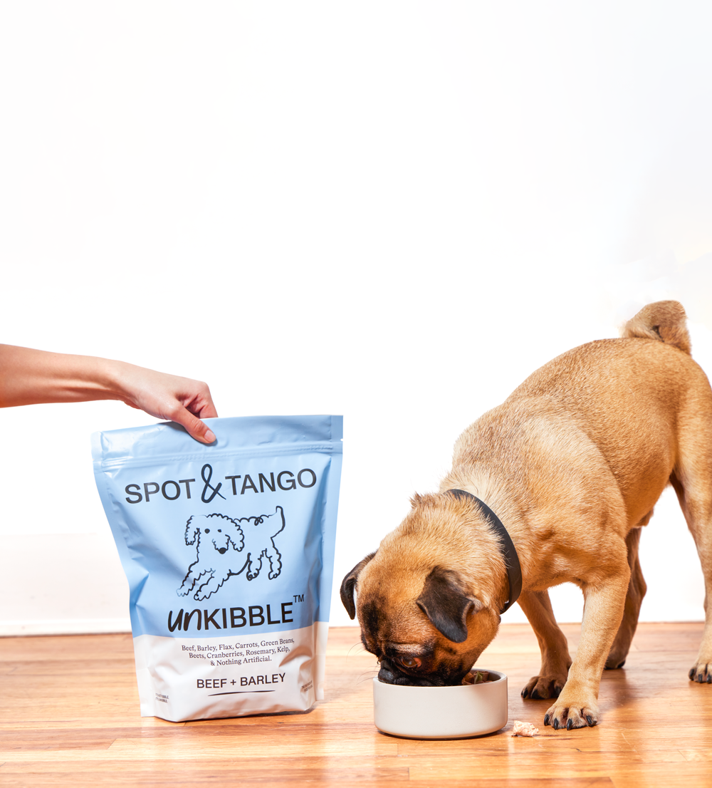
Spot & Tango
9.1Vet-formulated fresh trays and dehydrated UnKibble, portioned to the individual dog.

Lemonade Pet Insurance
9.4The lowest premiums in the category, with claims paid in minutes rather than weeks.

YETI Boomer 4 Dog Bowl
8.518/8 stainless, non-slip ring, dishwasher-safe — the last bowl you buy.
Recommendations are chosen by the Dr Murphies veterinary review board. Some links are affiliate links and we may earn a commission; this never affects our scoring, and brands do not see our copy before publication.
Dr Eleanor Murphy, BVSc
Founding editor and small-animal internist